Ultrasound
What is it?
Ultrasound is a safe and painless test. This technique produces images of the inside of the body using sound waves. It does not use radiation (X-rays).
The image is acquired by means of a probe (transducer) and a conductive gel that helps to better receive the signal. The high-frequency sound waves travel from the probe to the inside of the body. The organs and tissues return a sound (signal) that bounces back and is collected and processed in a computer to obtain the images.
These are obtained in real time and show the structure and movement of the internal organs, as well as the blood flowing through the blood vessels.
Studies performed:
- Heart and blood vessels, including the abdominal aorta and its main branches.
- Liver
- Gallbladder
- Spleen
- Pancreas
- Kidneys
- Bladder
- Uterus, ovaries
- Eyes
- Thyroid and parathyroid glands
- Scrotum (testes)
- Hepatic elastrography
- Breast
- Soft parts
- Muscular
Ultrasound is also used for interventional testing:
- Guiding procedures such as aspiration biopsies, where needles are used to sample cells from an abnormal area for laboratory testing.
- Obtain an image and guide the biopsy for breast, prostate,... studies.
- Inject contrast for the performance of ArthroMRI, ArthroCT tests.
How should I prepare?
Wear comfortable, loose-fitting clothing. You may need to remove all clothing and jewelry from the area to be examined.
You may be asked to wear a gown during the procedure.
Preparation will depend on the type of examination to be performed. Depending on the type of study you may be instructed not to eat or drink for up to 6 hours before your appointment, or to drink up to six glasses of water two hours before the test and to avoid urinating so that your bladder is full when the scan begins.
During the test the physician applies a small amount of gel to the area under examination and places the transducer there. The gel allows the sound waves to travel back and forth between the transducer and the area to be examined. The ultrasound image can be viewed immediately on a monitor.
Clinical Image Gallery:
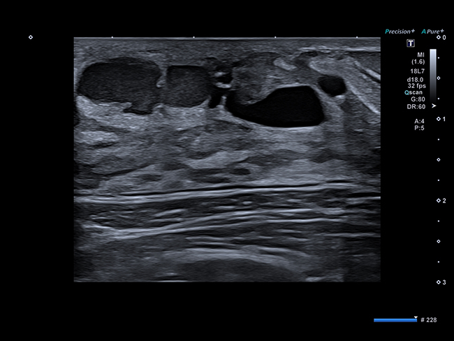
Dilated lactiferous duct

Subcostal short axis with tricuspid regurgitation on Color Doppler (left) and pulmonary regurgitation.

Kidney with SMI

Flexor tendon
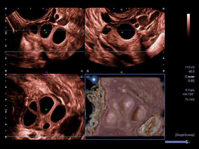
Ovary with follicles using Shadow Glass
